
Micrographs I Took with an Olympus Dissecting Microscope
After selecting the plant specimens and examining them under a compound Microscope, I placed them under an Olympus dissecting microscope which the friendly lab technician, Mr Mobius Leng, connected to a 29 inch CRT television for easier viewing.
-
I then took the micrographs with the dissecting microscope's built in digital camera, which stored the images on a 128MB SD card. I subsequently transferred the images to my IBM Thinkpad via USB2.0. I never knew microscopes had gone so high tech since my high school days!
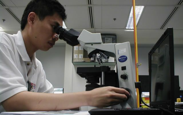
Working with my Olympus CX 31 Compound Microscope at the Plant Science Lab
.

Plant Specimens Sectioned with a Microtome, Biochemically Stabilized with 10% Formaldehyde in Phosphate Buffered Saline , Stained with Hematoxylin and Ready for Investigation
.
My Digital Micrographs Taken via Brightfield Microscopy with an Olympus Dissecting Microscope
While the Olympus CX31 compound microscope could magnify the tissue specimen up to 1000X (with oil), the dissecting microscope could only go up to 35X.
-
Dissecting microscopes generally allow larger, 3-D objects to be examined, as the lenses are positioned further away from the specimen. However, the drawback is that magnification is reduced. One advantage of the Olympus dissecting microscope I used was that it had a built in video capturing device, which fed moving images in real time to an external 29 inch television monitor. The dissecting microscope also had an SD card slot and a USB2.0 interface for fast and convenient transferring of images.
.
Micrograph of Fern Spores Taken Before Fine Focusing

Fern Spores at 15x Magnification (with fine focusing)

Fern Spores at 35x Magnification

Fern Spores at 15x Magnification (with fine focusing)
Fern Spores at 35x Magnification
Lichen at 10x Magnification (Overhead Lighting)

Lichen at 10x Magnification (Ambient Lighting)

Lichen at 10x Magnification (higher condenser setting)

Lichen at 10x Magnification (Condenser Deactivated)

Lichen at 10x Magnification (Side Lighting)

Corn Stem at 20x Magnification

Corn Stem at 35x Magnification

Onion Mitosis Root Tip at 20x Magnification

Closer view of Onion Mitosis Root Tip (35X)

Magnolia Stem Combination (20-35X)


Corn Stem (20-30X)


Moss (20-30x)


Pine Single Needle Leaf (20-30X)

Pollen at 15-35x Magnification


Silhoutte Lighting

(Bottom Lamp Dimmed)

posted by AL @ 11:22 PM
![]()
![]()
0 Comments:
Post a Comment
<< Home